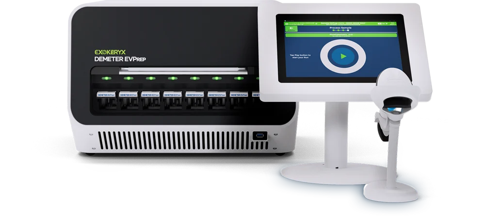
The science behind Demeter EVPrep
Demeter EVPrep leverages dielectrophoresis and the unique dipole moment of EVs to selectively isolate them from particles of the same size and density in biofluids. No more trading purity for yield.
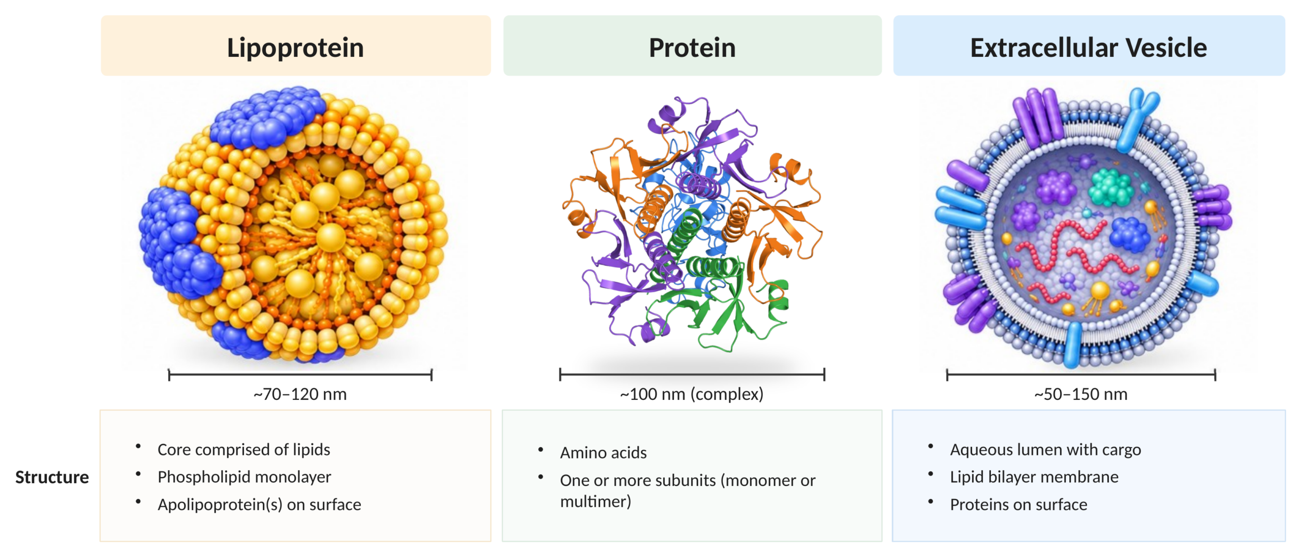
EVs have fundamentally different structures and composition
Biofluids and culture media are rich in proteins and lipoproteins that can share physical properties with EVs. Common techniques such as ultracentrifugation and size-exclusion chromatography use only density or size to separate EVs, leading to the co-isolation of contaminating material.
While EVs, proteins, and lipoproteins can overlap in size and density, these three groups have fundamentally distinct structures and compositions. EVs are particles with a protein-containing lipid bilayer membrane and an aqueous lumen containing cargo of nucleic acids, proteins and more. Our approach is to isolate EVs based on the electrical properties coming from their unique structure and composition.
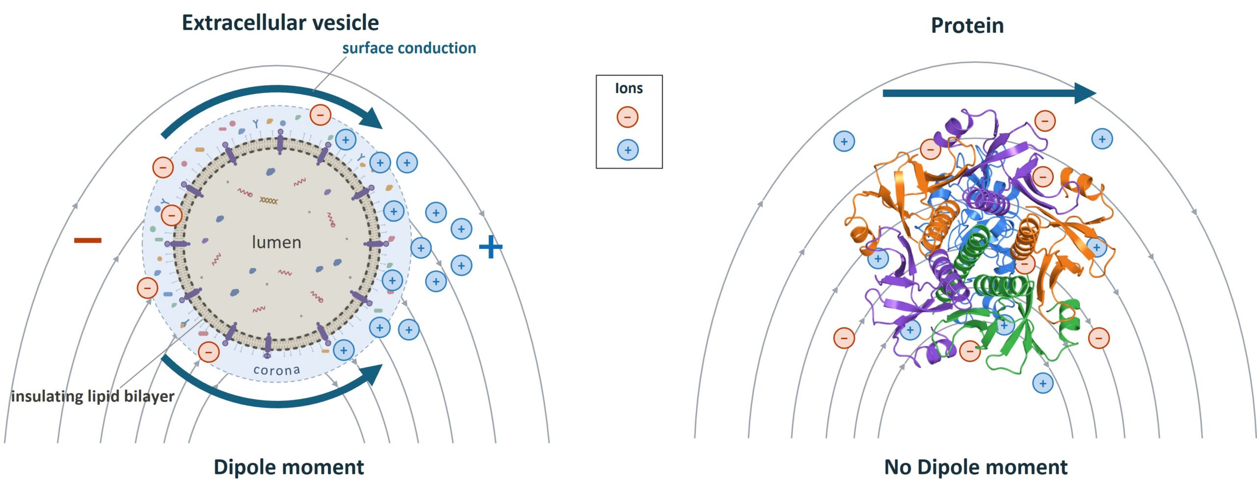
EV polarization under dielectrophoresis (DEP)
The lipid bilayer of EVs acts as an insulator to the conductive aqueous lumen. When an electric field is applied, the lipid bilayer causes the unbound charge to redistribute along the surface of the EV, polarizing it and creating a large dipole moment. Under the same conditions, proteins and lipoproteins do not experience the same charge redistribution and thus do not create a strong dipole moment. Therefore, the overall structure and composition, rather than the abundance of specific proteins in EVs, determines the dipole moment.
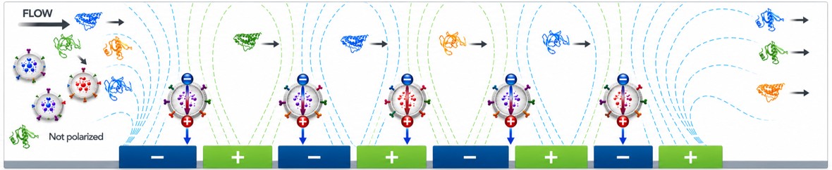
Polarized EVs are selectively captured
As the sample flows across the electrode array, polarized EVs are drawn to the electrode edges and held in place and other non-polarized proteins are washed away.
Fully automated DEP isolation of EVs in 3 steps
Polarize
EV-specific electric settings induce a large dipole moment in EVs.
Capture & Wash
EVs are held at the electrodes while non-polarized proteins are washed away.
Elute
Field off. EVs released into a small volume, concentrated and label-free.